
Master Course in Blepharoplasty (MCR)
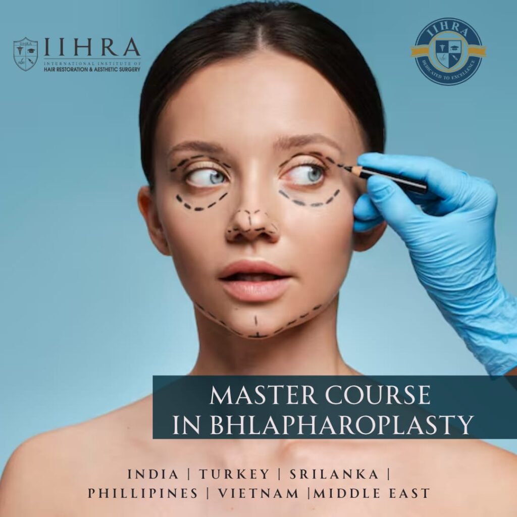
Comprehensive 2-Day Advanced Surgical & Hands-On Fellowship Program
- University Affiliation: Northwest International University (NIU) Affiliated Program
- Global Accreditation: Continuing Professional Development (CPD) Certified – UK CPD Approved
- Course Duration: 2 Days (Advanced Surgical Immersion)
- Instruction Format: Interactive Core Lectures, High-Definition Live Operating Room Demonstrations, & Supervised Hands-on Surgical Residency
- Minimum Eligibility: Strictly open to licensed Medical Practitioners specializing in Ophthalmology, Plastic Surgery, Otolaryngology (ENT), Maxillofacial Surgery, or equivalent advanced surgical disciplines.
This postgraduate-level surgical masterclass delivers an exhaustive, high-yield foundation in functional and cosmetic blepharoplasty, periorbital rejuvenation, and precision fat-pad manipulation. Led by an esteemed international faculty, the program transitions qualified surgeons from superficial injectables into the delicate, multi-layered surgical planes of the upper and lower eyelids.
Focus: Segmental Eyelid Anatomy, Upper & Lower Blepharoplasty Mechanics, and Vascular Safety Mapping
Module 1: Surgical Anatomy & Periorbital Tissue Layering
- Upper Eyelid Lamellae: High-resolution analysis of upper eyelid anatomy from skin to the orbicularis oculi muscle, orbital septum, pre-aponeurotic fat pads, levator aponeurosis, and Müller’s muscle.
- Lower Eyelid Lamellae & Fat Compartments: Detailed mapping of the medial, central, and lateral lower fat pads, the capsulopalpebral fascia, and the retaining ligaments (Orbicularis Retaining Ligament – ORL).
- Vascular & Nerve Architecture: Mapping the ophthalmic artery branches, angular vessels, and the course of the facial nerve motor branches alongside sensory infraorbital networks to avoid ischemic or paralytic events.
Module 2: The Blepharoplasty Consultation & Pre-Surgical Mapping
- Patient Selection & Dynamic Vectors: Differentiating between dermatochalasis, true steatoblepharon, and pseudoptosis. Identifying brow ptosis and performing the “snap-back” and “distraction” tests for lower lid laxity.
- Incision Geometry: Geometric design of the upper eyelid crease incision (preserving a minimum $20\text{ mm}$ vertical skin bridge) and lower lid subciliary vs. transconjunctival marking grids.
- Photographic Standards: High-resolution periorbital documentation, primary/secondary gaze tracking, and establishing legal medical consent document logs.
Module 3: Surgical Pharmacology, Local Blocks, & Tissue Dissection
- Ophthalmic Pharmacology: Formulation of tailored local anesthetic combinations (Lidocaine/Bupivacaine) with epinephrine, calculating maximum non-toxic dosages, and utilizing corneal shields safely.
- Upper Blepharoplasty Mechanics: Hydro-dissection techniques, skin-muscle flap excision strategies, targeted pre-aponeurotic fat resection, and conservative hemostasis.
- Lower Blepharoplasty Mechanics: Navigating the transconjunctival approach (for pure fat removal) vs. the transcutaneous subciliary approach (for skin/muscle laxity repair) and managing fat transposition matrices into the tear trough deformity.
Module 4: Cantal Stabilization, Closure, & Suture Rheology
- Canthopexy vs. Canthoplasty: Core principles of anchoring the lateral canthal tendon to the lateral orbital rim to prevent postoperative ectropion or lower lid malposition.
- Tension-Free Wound Closure: Skin-closure mechanics utilizing non-absorbable monofilaments, intradermal patterns, and edge-adaptation protocols to eliminate visible scarring.
Focus: Live Operating Room Case, High-Dose Complication Protocols, and Post-Op Continuity
Module 5: Pre-Operative Preparation & Theater Stabilization
- Clinical Protocol: Live patient marking with demographic instruments in the upright position, asymmetry mapping, ophthalmic surgical tray layout standardization, and establishing strict sterile fields.
Module 6: Live Surgical Execution – Case 1 (Four-Lid Blepharoplasty with Canthopexy)
- Upper Lid Execution: Supervised real-tissue skin-muscle excision, conservative fat debulking, and upper lid fold recreation under international faculty supervision.
- Lower Lid Execution: Transconjunctival or subciliary lower lid dissection, meticulous fat repositioning/excision, protective lateral canthopexy stabilization, and precision skin approximation.
Module 7: Comprehensive Complications Management & Emergency Protocols
- The Retrobulbar Hematoma: Acute recognition signs (proptosis, severe pain, loss of vision) and immediate execution of an emergency bedside Lateral Canthotomy and Cantholysis protocol.
- Lid Malposition Reversal: Algorithmic prevention and surgical revision pathways for postoperative lagophthalmos, ectropion, entropion, and scleral show.
- Soft-Tissue Corrosions: Reversing over-resection defects via secondary intent, skin grafting, or dynamic tissue suspension.
Module 8: Post-Surgical Continuity & Post-Op Maintenance
- The First 24–48 Hours: Suture care, cold-compress cooling matrices, lubricating ointment schedules, and strict activity limitations.
- Long-Term Periorbital Preservation: Formulating combination post-operative skincare regimes, energy-based periocular tightening, and targeted neuromodulation to protect surgical outcomes.
- Grand Final Review: Interactive clinical panel with the international surgical faculty, practice integration frameworks, and the distribution of Master Course Certifications from Northwest International University & UK CPD.